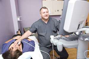
Ultrasound, or sonography, involves using high-frequency sound waves to produce pictures of the inside of the body. Ultrasound images are captured in real-time and show the structure and movement of the body’s internal organs, as well as blood flowing through blood vessels. This procedure is used to examine internal organs such as the heart and blood vessels, liver, gall bladder, pancreas, kidneys, bladder, uterus, ovaries, thyroid, and fetus in pregnant patients. Ultrasound is helpful in diagnosing a variety of conditions.
X-ray is simply a procedure used to evaluate injury to the extremities: arms, legs, hands, and feet. This type of imaging is frequently used to diagnose fractures or broken bones. Rockcastle Regional’s General Electric units provide the most advanced technology available.
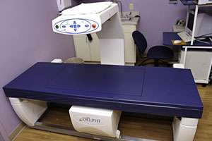
A bone density scan determines whether you have osteoporosis or are at risk of osteoporosis. Osteoporosis is a disease that causes bones to become more fragile and more likely to break.
In the past, osteoporosis could only be detected after you broke a bone. By that time, however, your bones could be quite weak. A bone density test makes it possible to know your risk of breaking bones before the fact.
A bone density test uses X-rays to measure how many grams of calcium and other bone minerals are packed into a segment of bone. A bone density test is a fairly accurate predictor of your risk of fracture.
The Seimens Magnatom Essenza, provides high-quality imaging for Rockcastle's radiologists, physicians, and patients. This MRI offers more diagnostic precision, while offering the patient a more comfortable exam.
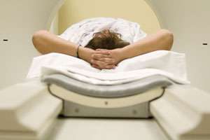
MRI (Magnetic Resonance Imaging) uses a powerful magnetic field and a computer to produce detailed pictures of organs, soft tissues, bone, and virtually all other internal body structures. This is a non-invasive, usually painless medical test that does not use radiation. Detailed images created by an MRI are better than those obtained by x-ray, ultrasound, or CT. Physicians use the MRI examination to help diagnose or monitor treatment for tumors of the chest, abdomen, or pelvis; lesions on the liver or other internal organs; tumors or abnormalities of the reproductive organs.
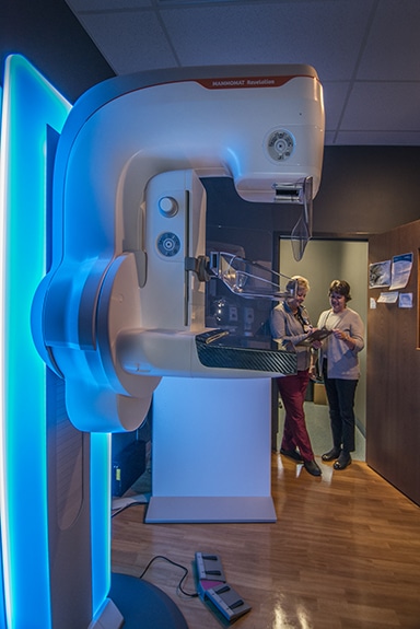
Rockcastle Regional offers the latest three-dimensional (3D) breast mammography technology. Each breast mammogram is read by a breast radiologist, a physician who specializes in reading x-rays of the breast.
Compression is key to getting a good mammogram. It prevents movement that can lead to blurred images. Compression spreads out the normal dense tissue of your breast, making it easier for breast radiologists to see through it and detect abnormalities. And, the more a breast is compressed, the less radiation exposure will be needed. At Rockcastle Regional, we know that compression is important, but it does not have to be painful anymore. Breast 3D mammograms are performed on the Siemens Healthineer’s MAMMOMAT Revelation mammography system because it makes comfort personal for each woman. Every woman is different, so our system automatically compresses only as long as the patient’s breast is soft and pliable. This means the patient gets exactly the right amount of compression for their breast type without losing any accuracy. The system also provides a wider-angle 3D technology for the highest-quality images for our breast radiologists to more easily and accurately detect small cancers. This means fewer false positives and callbacks for re-examination.
A mammogram is an x-ray of the breast. Mammograms can be used to check for breast cancer in women who have no signs or symptoms of the disease. This type of mammogram is called a screening mammogram. Screening mammograms usually involve two x-rays of each breast. They make it possible to detect tumors that cannot be felt. Screening mammograms can also find microcalcifications (tiny deposits of calcium) that sometimes indicate the presence of breast cancer.
Mammograms can also be used to check for breast cancer after a lump or other sign or symptom of breast cancer has been found. This type of mammogram is called a diagnostic mammogram. Signs of breast cancer may include pain, skin thickening, nipple discharge, or a change in breast size or shape. A diagnostic mammogram also may be used to evaluate changes found during a screening mammogram, or to view breast tissue when it is difficult to obtain a screening mammogram because of special circumstances, such as the presence of breast implants.
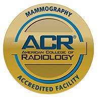
Rockcastle Regional has been awarded a three-year term of accreditation in mammography as the result of a recent review by the American College of Radiology (ACR). The ACR gold seal of accreditation represents the highest level of image quality and patient safety. It is awarded only to facilities meeting ACR Practice Guidelines and Technical Standards after a peer-review evaluation by board-certified physicians and medical physicists who are experts in the field.
Lung cancer is the number one cancer killer in the country. It is the most common cancer worldwide, representing 13 percent of all cancer diagnoses. Currently, there are three screening methods for lung cancer: sputum cytology (looks for cancer cells in a patient's sputum), standard chest x-ray, and low-dose CT scan. While all three methods detect signs of lung cancer, only studies on the low-dose CT scan show an acutal reduction in the likelihood of death from the disease. Additionally, the low-dose CT scan poses less risk to the patient than the regular diagnostic chest CT because the radiation exposure is much lower.
While low-dose CT scan is an excellent screening tool, it is not recommended for everyone. Those who are candidates for this test must meet all of the following "high-risk" criteria:
If you think you are a candidate for this screening, it is important to note that some insurance plans may not cover this test. It is best to check with your insurance plan for coverage of the screening and for any additional procedures, as there may be other costs associated even if the actual screening is free. The American Lung Association offers a checklist of questions to ask your insurance provider when calling to inquire about the screening.
Talk to you primary health care provider to discuss if a low-dose CT scan is right for you. The American Lung Association offers a helpful tool for you to know what to expect when you speak with your provider.
Nuclear Medicine is a subspecialty within the field of radiology that uses very small amounts of radioactive material to diagnose or treat disease and other abnormalities within the body. It is commonly used to evaluate organ function such as diagnostic studies of the heart and kidneys. Nuclear imaging procedures are noninvasive and usually painless medical tests that help physicians diagnose medical conditions. These imaging scans use radioactive materials called a radiopharmaceutical or radiotracer. Physicians use nuclear imaging to visualize the structure and function of an organ, tissue, bone, or system of the body.
© 2026 Rockcastle Regional Hospital. All rights reserved.
Website design + development by: